Basic Eye Anatomy
Understanding the anatomy of the eye is critical to understanding cataracts, how and why cataracts affect vision, and cataract surgery itself. Although the eye is small relative to most organs in the human body, it has many distinct anatomical parts, all of which contribute to the production of normal vision in one way or another. The following page explains basic anatomy of the human eye and highlights some structures in particular and how they relate to cataracts and cataract surgery.
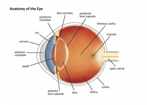
Figure 1.1: Normal Eye Anatomy
Eyelids and Lashes
The eyelids intermittently cover the front surface of the eye, forming a protective barrier. In addition, the eyelids help to replenish and spread the tear film that covers the front surface of the eye. The eyelashes also perform a protective function: they prevent dirt and other debris, as well as bacteria and viruses, from entering the eye, and are, therefore, very important in maintaining a healthy ocular surface.
Sclera
The sclera is the white substance that makes up the wall of the eye. It is very dense and strong. Normal sclera is white when we are born, but it may start to develop subtle changes in color as we age, ultimately taking on a yellowish hue.
Conjunctiva
The conjunctiva is the clear tissue that covers the front surface of the sclera, except in the location of the cornea. It functions in a manner similar to skin on the rest of the body. The conjunctiva serves as a protective barrier to entry for particles and organisms. In addition, it serves as a conduit for blood vessels and nerves and houses pockets of immune cells that help fight eye infections.
Pre-Corneal Tear Film
The pre-corneal tear film is the layer of tears that rest on the surface of the cornea. This tear film has three major components: water, fat and mucin. The health of each layer in the tear film is vital to the production of normal vision. This tear film is very important, as anyone who suffers from dry eye disease would tell you. Without an adequate tear film, your vision can be blurry and your eye can be uncomfortable.
Cornea
The cornea is the clear tissue that comprises the front “window” of the eye. It has several important functions. Its first and most important function is to refract light as it enters the eye. When we say a surface refracts light, we simply mean that it bends light in a certain way. Light rays can be bent toward one another, or away from one another, depending on the type of refracting surface. The cornea bends light rays toward one another, thereby helping the eye to focus the objects at which you look. Secondly, the cornea houses immune cells that help fight infections. Lastly, the inner layer of the cornea maintains the proper water content within the cornea itself. If this layer does not function properly, the cornea can become swollen and cloudy. In this case, it cannot perform its job of refracting light.
A clear and adequately functioning cornea is essential to normal vision. In the same manner that a dirty windshield or pair of glasses make your vision blurry, a cornea that is not clear will also reduce your vision.
The cornea is the location in which procedures such as laser vision correction (LASIK, LASEK, and PRK) and limbal relaxing incisions (LRIs) are performed. These are all procedures that help the eye to focus more clearly without glasses or contact lenses.LRIs are most commonly used in conjunction with cataract surgery, and will be discussed in greater detail in a subsequent chapter.
Anterior Chamber
The anterior chamber is a fluid-filled cavity situated between the cornea and the iris. It is filled with a nutrient-rich fluid called aqueous humor. This is the medium from which the front part of the eye derives many of its nutrients.
Iris
The iris is the structure that characterizes eye color. It is located approximately three millimeters behind the cornea. The central opening in the iris is called the pupil, and acts as an aperture. By increasing and decreasing in size, it controls the amount of light that enters the eye. This is the structure that must be dilated in order to adequately visualize the lens, retina and optic nerve in the back of the eye. It must also be dilated in order for cataract surgery to be performed.
Posterior Chamber
The posterior chamber is the fluid-filled cavity situated between the iris and the lens. It is also filled with aqueous humor. It is a very tiny space in which a cataract surgeon works to remove a cataract.
Crystalline Lens (The Lens)
A normal crystalline lens is clear at birth. It functions as the second refracting surface in the eye (remember, the cornea is the first refracting surface). Just like the cornea or the lenses in your glasses, it must be clear in order to achieve normal vision. By changing its shape, the lens allows for accommodation to occur. Accommodation is the process of focusing from distance to near, and at all distances in between. The eye loses its ability to accommodate as we age, which is why most people develop the need for reading glasses around the age of 40.
Capsular Bag and Lens Zonules
The lens capsule, or the capsular bag, is the structure that holds the lens in a central position within the eye. It is composed of an anterior and posterior capsule. Attached to the periphery of the capsule are tiny string-like structures called zonules. These are microscopic fibers that connect the capsule to the ciliary muscles along the inside wall of the eye. They are important structures that help maintain the appropriate orientation of the lens inside the eye, and allow for the process of accommodation to occur.
Ciliary Muscles
The ciliary muscles are tiny muscles that cause the shape of the lens to change as they contract and relax. This is the process of accommodation, which allows the eye to change its point of focus.
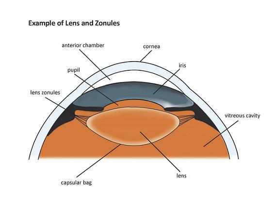
Figure 1.2: Illustration showing normal crystalline lens and zonules.
Vitreous Humor
The vitreous humor, or vitreous, is a dense jelly-like substance that fills the space within the eye between the lens and the retina, called the vitreous cavity.
Retina
The retina covers the inside back wall of the eye. It contains cells that are activated by light and transmit a signal to the optic nerve in order to produce vision. The center of the retina is called the macula. This is where the images of objects in your central vision are projected. It is also the structure that is affected in macular degeneration. The retina is commonly referred to as “the film in the camera,” because this is where the visual image is received and imprinted, similar to the process that happens when a picture is taken with a camera.
Optic Nerve
This is the structure that carries visual information that has been received by the retina from the eye to the brain. It is vital to the formation of normal vision. This is also the structure that is damaged in glaucoma.
These structures all work together in a remarkable display of teamwork to produce normal vision. Damage to or destruction of any one of these anatomical parts can lead to a decrease in vision. The following chapter will discuss how vision works in the human eye. Understanding how vision works will help you to understand the impact of cataract formation on vision, and how its treatment can be so effective at restoring vision.
© Vision Information Services, LLC, Mooresville, NC 2012